

Calcaneal apophysitis, commonly referred to as Sever’s disease, is a frequent cause of heel pain in physically active children and adolescents. The condition was first named in the early 20th century by James Warren Sever, MD, an American orthopedist. Despite the name, Sever’s is not a true disease; it is neither infectious nor degenerative and does resolve with time.
It is a painful overuse injury related to skeletal immaturity. It occurs at the calcaneal apophysis, located at the posterior aspect of the heel bone, where the Achilles tendon inserts. Sever’s disease is an inflammatory response and mechanical stress reaction at the growth plate of the heel during periods of rapid growth. It is one of the most common musculoskeletal complaints seen in young podiatry patients and pediatric sports medicine.
Etiology
In anatomy, the term physis refers to a growth plate, a cartilaginous area where bone growth occurs. The physis allows the bone to lengthen before fusing and taking its final shape in adulthood. Hence, apophysitis is an inflammation or stress injury around a growth plate, where muscle attaches to the bone nodule. This area at the growth plate is susceptible because it is mechanically weaker than the surrounding bone. Apophysitis can occur in many parts of the body when there is overuse or excessive strain. Besides the heel, other common areas of complaint include the knee (Osgood-Schlatter’s disease), the hip, and the elbow.
The calcaneal physis typically fuses by the time adolescents are approximately 14-16 years old, though this varies based on gender and individual growth patterns. During adolescent growth spurts, bones often lengthen more rapidly than muscles and tendons can adapt, putting them under increased tension. In the case of Sever’s, a sudden elongation of the tibia can result in the Achilles tendon becoming tight, which increases traction on the posterior calcaneal apophysis. If a child is very active, the heightened repetitive forces can cause microtrauma at the growth plate.
Contributing Factors
There are several documented environmental and biomechanical factors that contribute to the development of calcaneal apophysitis. A rapid growth spurt can be a trigger, especially if it coincides with intense training or a sudden increase in physical activity such as joining a team or returning to athletic practice for the new season. The hard surfaces of running tracks and sports courts that lack impact absorption can exacerbate the condition.
Biomechanically, any limit in ankle range of motion can put excess strain on the Achilles tendon. Sometimes a pediatric flatfoot is the result of a short gastroc-soleus, and a child’s apparent pes planus is due to the restricted range of motion at the ankle. A leg length discrepancy may also be the culprit, where a patient reflexively accommodates for a limb length inequality through compensations in the foot and leg; and likewise for any asymmetrical limb segment rotations or joint motions. Finally, increased body weight is a factor as it raises load and ground reaction forces.
Treatment
Once the diagnosis is confirmed, the first and obvious step to treatment, especially when the pain level is high, is an immediate cessation or at least a drastic reduction in physical activity. Rest may not be welcome, but it will reduce traction on the Achilles and is necessary for healing to occur. Icing also helps. The second step is to reduce the mechanical load by avoiding running, jumping, etc. during the acute phase. Sometimes a light schedule of cross-training such as swimming or biking can be suggested as alternatives for staying in shape.
Stretching is an important element of the rehabilitation process and should be supervised by a physical therapist. The goal is to stretch the posterior muscles and tendons without putting further strain on the growth plate. Gentle stretches and muscle massaging performed daily will help improve flexibility in the lower limb. Taping and compression may also be recommended.
Lifts, Shoes, and Orthotics
Strain on the physis can be lessened with the use of heel lifts. Elevating both heels by 3-5 mm can reduce the mechanical pull and help the pain subside. Cork heel raises, silicone cushions, or rubber heel cups are all used successfully. Unless there is a noted leg length difference, heel lifts, cushions, and cups must always be dispensed bilaterally to avoid creating any imbalance.
Footwear is an important consideration as it provides cushioning and control for the foot. At a minimum, the shoe and/or the insole should have a base level of shock absorption to reduce the effect of impact forces. The sole should easily flex at the ball, along with firm heel counters to control rearfoot motion. There should be some amount of heel elevation or drop from the heel to the ball. Soccer cleats are a good example of poor footwear as they tend to be hard and flat. As a remedy, adding heel cushions into the narrow boots both elevates the heel and provides some shock absorption. Likewise, minimalist footwear, flat sneakers, or going barefoot should be avoided both on and off the field. And all pediatric footwear should be properly sized to allow for growth in the young, developing foot.
Orthotics are helpful when biomechanics are thought to be a contributing factor. For instance, raising the medial longitudinal arch and using a deep heel cup (> 20 mm) to control rearfoot motion can be beneficial for a child with correctable feet. The orthotics do not need to be custom; there are several brands of good orthopedic prefabs that can be modified. It is possible to take a semi-rigid thermoplastic shell and add various components such as bilateral heel raises, heel cushions, arch pads, and EVA top covers.
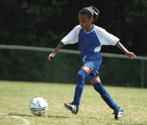

- Patients with calcaneal apophysitis tend to fit a certain profile.1 Usually they are eight to 15 years old, with an average age of ten to 11. Although not uncommon in girls, it is more frequent in boys. It can be reported bilaterally or unilaterally. The patients are often highly active adolescents engaged in soccer, basketball, running, gymnastics, and other demanding disciplines.
- The typical symptom is heel pain worsened by activity and relieved with rest. It differs from plantar fasciitis as the pain will be localized to the posterior or plantar aspect of the heel, and plantar fasciitis pain can lessen with activity. Usually, the onset of the heel pain has been gradual with no recall of a specific traumatic event or injury. It is not unusual to note limping and pain during or after sports or having stiffness in the morning or at the start of activity.
- Physicians diagnose the condition by physical examination.2 Most patients have tenderness at the posterior calcaneal apophysis or bulge, and they will often report pain during a calcaneal squeeze test. Imaging is not usually ordered because the area around the immature calcaneal physis tends to be unclear. Physicians will rule out other possible diagnoses such as a calcaneal fracture or a nerve impingement.
- The good news for patients is that calcaneal apophysitis will resolve once the growth plate matures and fuses. Additionally, when managed properly it does not cause long-term disability or structural heel damage. However, young athletes are often eager to return to practice quickly and get frustrated when asked to slow down or sit it out.
Symptoms can last weeks to several months, but it is important to be cautious in returning to full intensity activity as recurrences happen. Some children experience intermittent symptoms over one to two years during successive growth spurts. Recognizing and addressing the condition early on can be helpful in managing the condition.
Conclusion
Calcaneal apophysitis is a common cause of heel pain in growing children and adolescents as the result of repetitive mechanical stress at the calcaneal growth plate. It typically appears during periods of rapid growth, particularly in active children and is best addressed early by quickly modifying strenuous activity. Proven treatment protocols include stretching and physical therapy, proper footwear selection, the use of bilateral heel lifts, and foot orthotics when necessary3. With appropriate management and a little patience, nearly all patients return to full activity.
Séamus Kennedy, BEng (Mech), CPed, FAAOP(A), is president and co-owner of Hersco Ortho Labs, New York. He can be contacted at seamus@hersco.com or by visiting www.hersco.com.
References
- Valmassy, R. L. 1996. Clinical Biomechanics of the Lower Extremity Mosby.
- Alexander, I. J. 1990. The Foot: Examination and Diagnosis Churchill Liningstone.
- Hernandez-Lucas, P. and R. Leirós-Rodríguez, et al. 2024. Conservative treatment of Sever’s Disease: A systematic review. Journal of Clinical Medicine 13(5):1391.
Teen Soccer Player: Isaiah Love/stock.adobe.com.