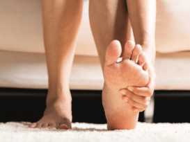
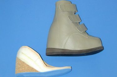
Adult-acquired flatfoot (AAF) is defined as a progressive deterioration of the static and dynamic structures that support the foot. Although frequently linked with posterior tibial tendon dysfunction (PTTD), AAF is a far broader syndrome that describes a gradual but painful breakdown of the weight bearing foot.

In a previous article, “Understanding Adult-Acquired Flatfoot” (The O&P EDGE, November 2014), we looked at some of the causes of this condition, which usually begins with bilateral pes planus, progresses to PTTD, and can ultimately end up as a fixed deformity with midfoot collapse and forefoot abduction. Along this trajectory, the foot moves through clearly defined stages as the key supporting ligaments are attenuated, midfoot joints become subluxed, and the foot becomes more rigid.

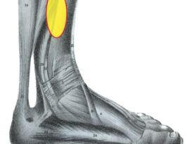
Several tests can be used to evaluate and stage AAF. The patient’s initial complaint will often be unilateral pain along the course of the posterior tibial (PT) tendon. It is helpful to recognize that, although the symptom may appear to be local, the effect on foot function is occurring in all three planes. For example, a lower arch due to PT muscle weakness can lead to a restriction of the critical first metatarsal phalangeal joint (MPJ) (sagittal plane motion). This, in turn, blocks the windlass mechanism, resulting in an unstable midfoot after heel lift, which is often seen as calcaneal eversion (frontal plane motion). Although not an exhaustive list, the spectrum of tests in this article will assess the loss of foot strength and function in several dimensions. Radiographic analysis and nerve conduction tests will confirm the extent of the condition.

Given that AAF progresses in a somewhat defined pattern, there are appropriate treatment protocols to address each stage of the condition. The goal is to provide adequate support to prevent further pathology-without restricting beneficial motion. Thus, patients who exhibit the early stages of AAF can often be treated successfully with functional foot orthotics and physical therapy, whereas patients in the later stages require the more restrictive control of AFOs and perhaps surgery.
Patients with AAF
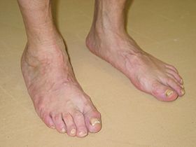
Patients with AAF typically present with bilateral flatfoot deformities.
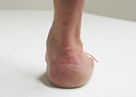
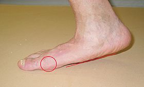
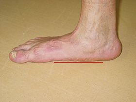
asymmetrically lower arch
Usually there will be an asymmetrically lower arch on the affected side. Here we see the complete absence of the medial longitudinal arch (MLA).
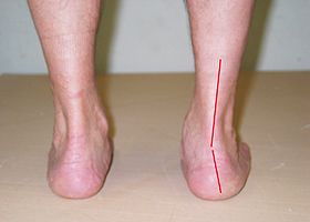
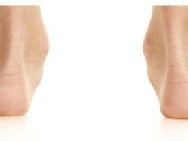
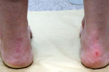
calcaneal valgus
In the frontal plane, note the eversion and calcaneal valgus.
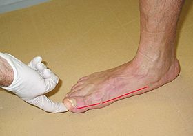
abduction of the forefoot
Often there will be an abduction of the forefoot, also referred to as the “too many toes” sign, indicating a transverse plane component to the deformity.
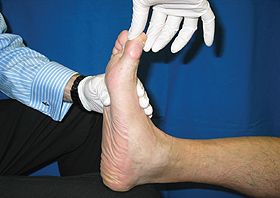
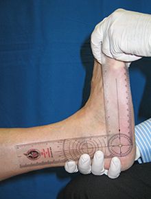
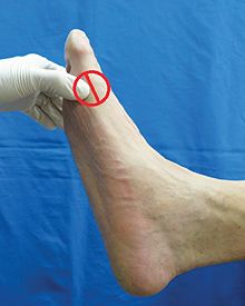
Test the range of motion
Test the range of motion (ROM) of the first MPJ in a non-weight bearing position. In a normal foot, the hallux should dorsiflex at least 50 degrees. Smooth motion of the first MPJ is essential to foot function in the sagittal plane.
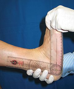
Perform the Hubscher maneuver
Perform the Hubscher maneuver on the weight bearing foot to test the functional ROM. Less than 12 degrees of dorsiflexion indicates a functional hallux limitus. If motion is restricted, then the windlass mechanism is blocked. The windlass mechanism is essential for pulling the plantar fascia taut, stabilizing the midtarsal joint during the propulsive phase of gait, and rotating the lower leg externally.
Use the Silfverskiold test
Use the Silfverskiold test to check for available ROM at the ankle. Less than 10 degrees of ankle dorsiflexion represents a restriction and some level of equinus.
dorsiflexion of the foot available
If there is increased dorsiflexion of the foot available with the knee flexed, it indicates a tight gastrocnemius. The presence of equinus places undue stress on the support structures of the midfoot, leading to their gradual deterioration.
positive single-foot heel rise test
In a positive single-foot heel rise test the patient will perform the test with difficulty, and in the later stages cannot complete the test at all. Note the lack of motion at the first MPJ and the non-development of the MLA. Seen from the posterior, the calcaneus will fail to invert.
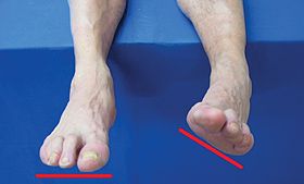
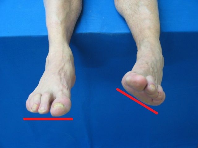
supination lag test
The supination lag test requires the patient to sit with both legs suspended and then attempt to have the soles of the feet face each other. It tests the ability to invert and adduct when non-weight bearing. In the presence of AAF, where muscles and ligaments have been disrupted, there will be an obvious asymmetry. With rupture of the PT tendon, the foot cannot cross the midline of the tibia.
Test the strength of the PT muscle
Test the strength of the PT muscle by pressing and abducting the lower 50 percent of the first metatarsal head. The ankle should hang plantarflexed, and the patient is asked to push (rotate) his or her foot inward (adducting) against the examiner’s thumb, while the examiner’s other hand stabilizes the lower leg. Often there will be noted weakness on the affected side compared to the contralateral side.
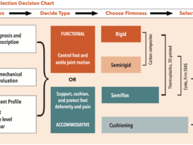
Select the orthotic device
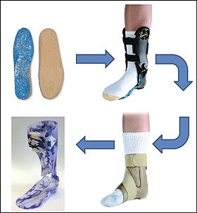
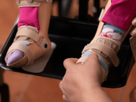
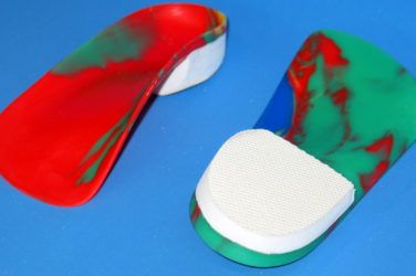
Select the orthotic device based on the patient’s current condition: It should offer good support and control without being overly restricting. As AAF progresses, the effects of the condition are seen in all three planes so the orthosis must respond accordingly, providing appropriate control and support in all planes without excess restriction. Foot orthotics and short, articulating AFOs used in conjunction with supportive, extra-depth shoes work well in the early stages of AAF. Gauntlets and AFOs provide more support, limiting motion above and below the ankle.

For a more complete review of the etiology and staging of AAF, read “Biomechanics and Clinical Analysis of the Adult Acquired Flatfoot,” Douglas Richie Jr., DPM, FACFAS, Clinics in Podiatric Medicine & Surgery, 2007.
You May Also Like

The Diabetic Epidemic

The statistics about diabetes are staggering. In 2017, the Centers for Disease Control and ...

Preventing Running Injuries

Running has been referred to as the king of all workouts. It is an ...

Mechanical Knee Pain

The knee is the largest and strongest joint in the body, consisting of the ...

Leg Length Discrepancy

Leg length discrepancy (LLD) is a diagnosis that appears deceptively simple, one limb being ...

Achilles Tendinopathy

In Greek mythology, Achilles’ mother, Thetis, dipped her young son headfirst into the river Styx ...